

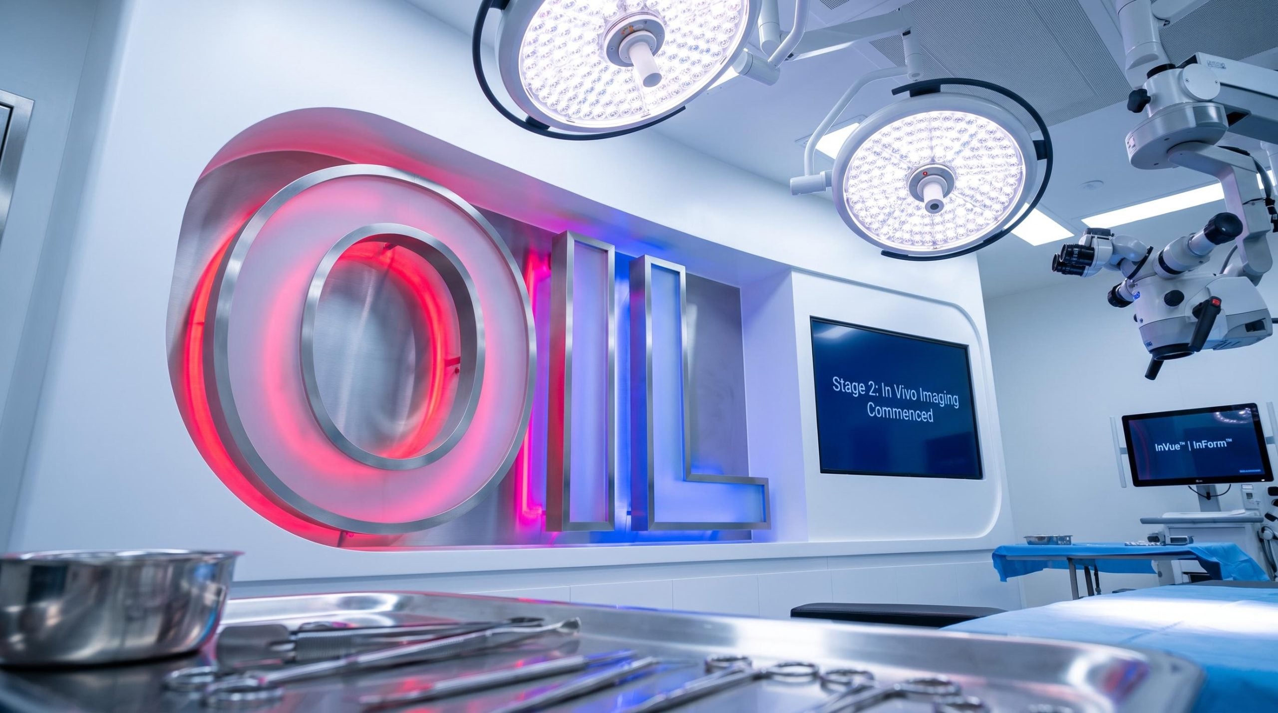
Optiscan advances head and neck cancer study to Stage 2 ahead of schedule
Optiscan Imaging (ASX: OIL) has advanced its first-in-human head and neck cancer imaging study to Stage 2 ahead of schedule, marking progress at Perth’s St John of God Murdoch Hospital. The study utilises the company’s Optiscan InVue™ precision surgery platform for real-time intraoperative imaging and the Optiscan InForm™ digital pathology platform.
The quicker-than-expected transition delivers time and cost efficiency benefits, while accelerating clinical validation, FDA regulatory evidence generation and commercial readiness.
Advancement from Stage 1 ex vivo imaging was approved by the SJG Murdoch Human Research Ethics Committee, a decision based on the rate, quality and variety of tissues imaged to date. Notably, this is the first human clinical study to image head and neck cancer tissues in vivo during surgery.
When big ASX news breaks, our subscribers know first
What Stage 2 involves and why the acceleration matters
Stage 2 focuses on obtaining in vivo images during surgery using Optiscan InVue™, supplemented by ex vivo imaging using the Optiscan InForm™ device. This follows Stage 1, which centred on ex vivo imaging, with the advancement approved on the strength of accumulated data.
The study, initiated late last year (ASX announcement dated 3 December 2025), is led by Perth-based head and neck cancer surgeon Dr Chady Sader, who assisted in recruiting the first 10 cases during Stage 1. Imaging from that initial set supported the accelerated transition.
The Stage 2 advancement builds on the foundation established when Optiscan initiated its 50-patient head and neck cancer study in late 2025, a program designed to generate FDA-quality data while simultaneously activating partnerships with Australian Clinical Labs and Long Grove Pharmaceuticals.
Australian Clinical Labs continues to support the study through pathology specimen analysis using the InForm™ platform across both fresh and fixed tissue samples. Leveraging previously announced collaboration agreements with Australian Clinical Labs and Long Grove Pharmaceuticals, the study utilises sodium fluorescein as the contrast agent in combination with the InVue™ device.
Key facts for Stage 2 include:
- Site: St John of God Murdoch Hospital, Perth
- Lead investigator: Dr Chady Sader
- Pathology partner: Australian Clinical Labs (InForm™)
- Contrast agent: sodium fluorescein (with InVue™)
- Purpose: real-time intraoperative imaging and oral cancer AI algorithm development
Why real-time imaging matters for cancer surgery
Under the current standard of care, surgical margins are assessed post-operatively through pathology review. This means close or positive margins are identified only after surgery has concluded. That delay can drive higher recurrence risk and treatment escalation.
Real-time cellular imaging with Optiscan InVue™ is designed to enable immediate intraoperative margin assessment during the procedure itself. According to the company, this could support improved margin clearance and reduced recurrence risk.
Optiscan’s core technology relies on miniaturised digital endomicroscopes, small imaging devices that produce single-cell-level images. The company states the spatial resolution is more than 1000x that of medical CT and MRI scans.
For investors, the ability to bridge the gap between surgery and pathology addresses a substantial clinical need and market opportunity across surgical oncology.
| Workflow stage | Current standard of care | Standard of care with InVue™ |
|---|---|---|
| Imaging timing | Post-operative pathology review | Real-time cellular imaging during surgery |
| Margin assessment | Close/positive margins identified after surgery | Immediate intraoperative margin assessment |
| Clinical outcome | Higher recurrence risk and treatment escalation | Improved margin clearance and reduced recurrence risk |
Clinical, regulatory and commercial significance
Clinical significance
Stage 2 enables live intraoperative imaging during head and neck cancer surgery, supporting assessment of tissue architecture and surgical margins in procedures where real-time decision-making is clinically important.
Regulatory significance
The study will generate paired imaging and pathology evidence across fresh, fixed and in vivo tissue settings. This strengthens the dataset intended to support Optiscan’s US FDA submissions and future US clinical study design.
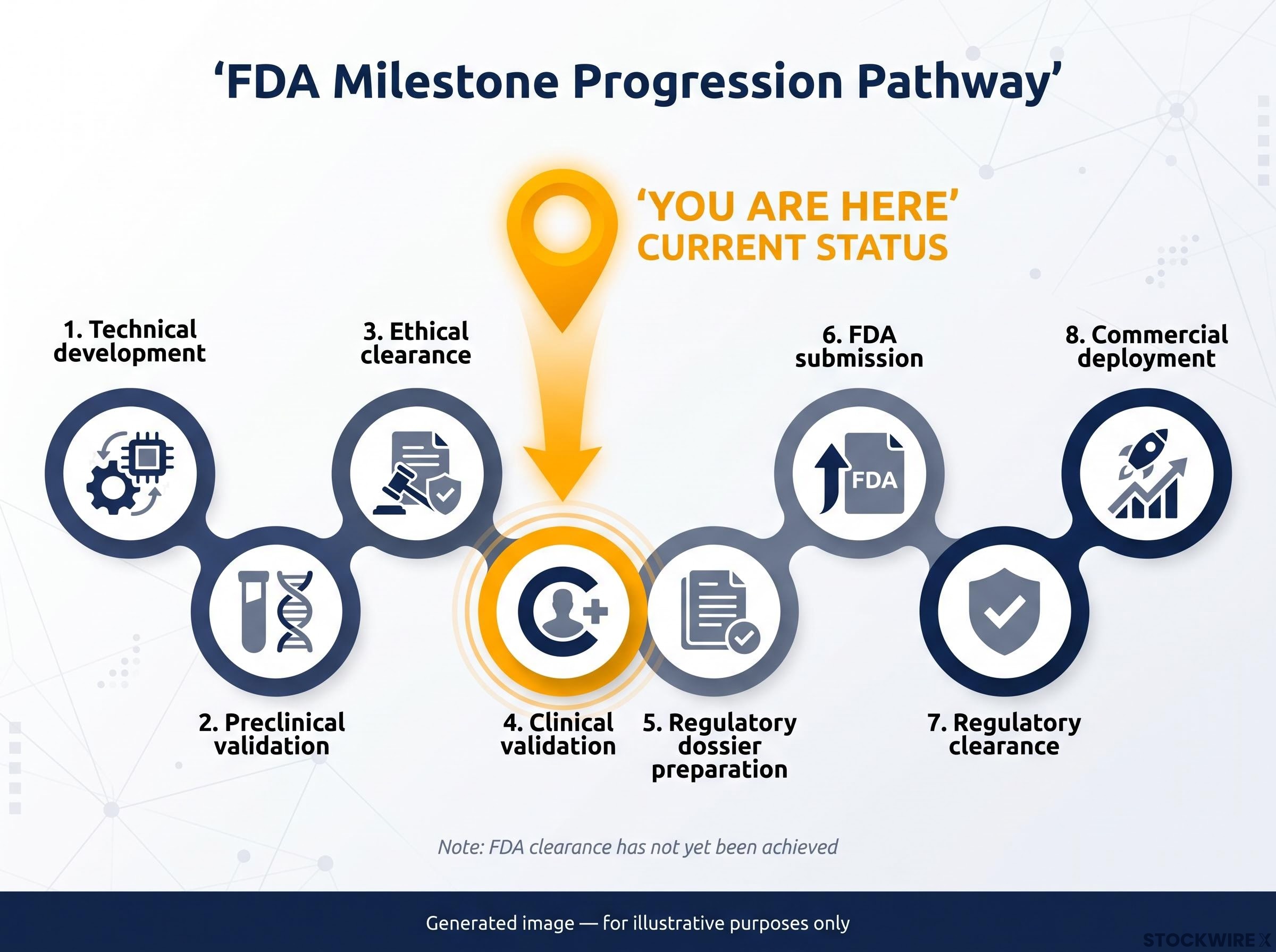
The company’s FDA milestone progression spans technical development, preclinical validation, ethical clearance and clinical validation, where Optiscan currently sits, followed by regulatory dossier preparation, FDA submission, regulatory clearance and commercial deployment. FDA clearance has not yet been achieved.
Optiscan is running parallel clinical programs across cancer types, and its breast cancer imaging study reached the halfway recruitment mark in June 2026 with 25 of 50 patients imaged at The Royal Melbourne Hospital, zero adverse events recorded, and FDA submissions for InVue and InForm devices on track for H2 CY2026.
Commercial significance
Demonstrating workflow integration in a complex surgical oncology setting supports future adoption of Optiscan InVue™ and Optiscan InForm™ across precision surgery, digital pathology and broader cancer imaging applications.
For investors, the study represents data compounding toward the FDA pathway while feeding multiple strategic priorities, including AI development, future US clinical activities and regulatory progression.
Management perspective and what comes next
Dr Camile Farah, CEO and Managing Director
“We are thrilled to see Stage 2 of the SJOG study utilising our InVue™ and InForm™ platforms commence ahead of initial expectations. This achievement is a testament to the ease of integration of both these innovative platforms into surgical and pathology workflows, as well as the strength of the collaboration between Optiscan, SJOG, and Australian Clinical Labs.”
Dr Farah noted the study is helping validate the platform across multiple tissue types, procedures and healthcare settings, with data expected to contribute to strategic priorities spanning AI development, future US clinical activities and regulatory pathways.
Dr Chady Sader, Lead Investigator, SJG Study
“I am encouraged by the rate of recruitment, the quality of images produced to date and their concordance with standard histopathology. Progression to live imaging is an important next step in evaluating how this technology may assist surgeons during head and neck cancer procedures.”
Data from the study feeds three key priorities: development of the oral cancer AI imaging algorithm, upcoming US FDA regulatory submissions collating data from multiple studies, and future US clinical activities. No specific FDA submission date has been disclosed.
Optiscan describes itself as a commercial stage medical technology company developing digital pathology and precision surgery solutions that enable live optical biopsy across life sciences, diagnostic and surgical applications.
Don’t Miss the Next Healthcare Breakthrough on ASX
Big News Blast delivers FREE breaking ASX healthcare news directly to your inbox within minutes of release, complete with in-depth analysis already done for you. Join 20,000+ investors who stay ahead of market-moving announcements before the broader market reacts. Click the “Free Alerts” button at StockWire X to start receiving alerts today.