Micro-X Achieves World-First Human Brain CT Images at Royal Melbourne Hospital
World-first human brain images mark a defining moment for Micro-X’s Head CT program
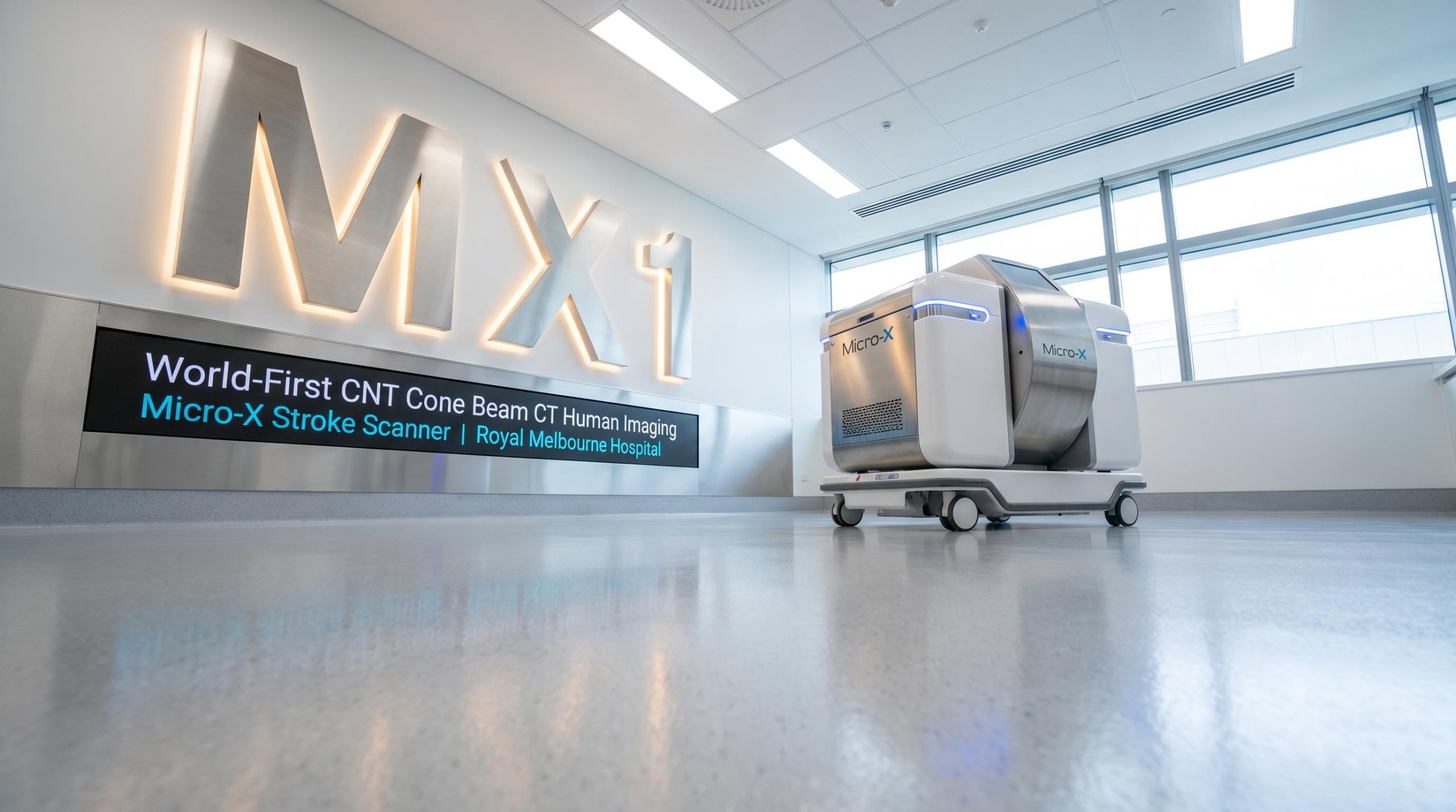
Micro-X Ltd (ASX: MX1) has achieved a world-first in medical imaging, acquiring CNT Cone Beam CT human patient images using its Head CT scanner at Royal Melbourne Hospital. The milestone marks the formal commencement of the Micro-X Head CT human imaging study, capping four years of development and transitioning the programme from bench-level testing to real clinical data collection.
The study, formally titled the Micro-X Stroke Scanner multi-centre pilot, will enrol 108 patients across four brain pathology conditions: ischaemic stroke, intracranial haemorrhage, brain tumour, and stroke mimic. The full enrolment period is estimated to take six to nine months from commencement.
Anthony Skeats, Chief Operating Officer
“This milestone represents four years of relentless innovation, belief and determination from our team. Achieving first human imaging represents a significant technical milestone for the Company and for cold cathode emitter technology. We look forward to completing the pilot study over the coming months to generate the data required to inform our regulatory pathway.”
Key announcement takeaways:
- World-first CNT Cone Beam CT human patient images acquired at Royal Melbourne Hospital
- Initial real patient datasets to be used to refine imaging algorithms
- $0.4M MRFF milestone payment triggered following completion of the second test bench
When big ASX news breaks, our subscribers know first
Inside the Head CT: what the technology does and why it matters
Conventional X-ray systems rely on heated filaments to generate electrons, a process that demands significant energy and produces bulky hardware. Micro-X’s Head CT uses cold cathode emitter technology based on carbon nanotube (CNT) sources, which emit electrons without requiring heat. This fundamental difference allows for a substantial reduction in size, weight, and power consumption compared to traditional X-ray tubes.
The scanner incorporates a curved array of 21 electronically controlled CNT X-ray tubes with fast electronic switching capability. This array, combined with proprietary reconstruction software, generates three-dimensional brain images. The result is a device designed to fit inside a standard road ambulance and weigh under 70kg. Current testing indicates an effective patient dose of ≤0.75 mSv, which is substantially lower than the dose associated with standard brain CT imaging.
From hospital to ambulance: the stroke care gap this device is designed to close
In pre-hospital settings, paramedics responding to a suspected stroke cannot currently distinguish between the two primary stroke types: ischaemic stroke (caused by a blockage) and intracranial haemorrhage (caused by bleeding). This distinction is critical because the front-line treatment for ischaemic stroke, thrombolysis (clot-dissolving medication), is contraindicated in haemorrhagic stroke. Without access to CT imaging in the field, treatment decisions are delayed until the patient reaches hospital.
The Head CT is purpose-built to address this gap. Dr Anna Balabanski, lead clinical researcher and neurologist with the Australian Stroke Alliance, described the clinical ambition clearly:
Dr Anna Balabanski, Australian Stroke Alliance
“The commencement of human imaging marks an important milestone in evaluating this novel technology. The pilot study will allow us to assess image quality and clinical utility against conventional CT in a controlled hospital environment. The Australian Stroke Alliance is now keen to see the technology evolve beyond a hospital-based scanner into a truly portable device, capable of reaching patients wherever they are.”
Milestone payments, study structure, and the road to regulatory submission
Second test bench triggers $0.4M MRFF payment
Alongside the commencement of human imaging, Micro-X has completed the manufacture of a second Head CT test bench intended for the second trial site at Royal Adelaide Hospital, including safety and verification testing. This completion satisfies Milestones 6b and 7b of the Medical Research Future Fund (MRFF) programme with the Australian Stroke Alliance and triggers a $0.4M payment to Micro-X.
Clinical study at a glance
| Study Element | Detail |
|---|---|
| Study title | The Micro-X Stroke Scanner: A prospective multi-centre pilot study of a novel miniaturised cone beam computed tomography scanner for identifying neuroimaging-based contraindications to stroke thrombolysis in hospitalised patients |
| Indication | Patients with (1) ischaemic stroke, (2) intracranial haemorrhage, (3) brain tumour, or (4) stroke mimic |
| Primary objective | To investigate the ability of the scanner to identify neuroimaging-based contraindications to stroke thrombolysis and assess clinical utility for acute stroke assessment |
| Study design | Prospective multi-centre non-randomised single group within participant cross-over pilot comparative study |
| Sample size | 108 patients |
| Duration | Estimated 6-month enrolment period followed by analysis and reporting |
Government funding backing the programme
The Head CT programme has received $8M in funding from the Australian Government’s Medical Research Future Fund (MRFF), with a further $4.4M committed through the Australian Government Industry Growth Programme. The depth of government investment reflects the programme’s credibility and the clinical problem it is targeting. Key collaborators include the Australian Stroke Alliance, Monash University’s Design Health Collab, and Johns Hopkins University.
The next major ASX story will hit our subscribers first
What investors should watch from here
With human imaging now confirmed, investors should monitor the following near-term catalysts and milestones as stated in the announcement:
- Algorithm refinement phase commences — initial real patient datasets are now being applied to refine imaging algorithms
- Study completion expected within 6–9 months — full enrolment of 108 patients across all four target conditions, followed by analysis and results publication
- Reader study completion required — images cannot be released publicly until the reader study is complete, to preserve the integrity of trial results
- Regulatory pathway informed by pilot outcomes — management has indicated the study data will directly guide the regulatory strategy
- Royal Adelaide Hospital activation — the second test bench is ready, and the second trial site is positioned to contribute to the study as it scales
The commencement of human imaging is a material de-risking event, moving the Head CT from development-stage asset to active clinical validation.
Don’t Miss the Next Healthcare Breakthrough on ASX
Big News Blast delivers FREE breaking ASX healthcare news straight to your inbox within minutes of release, complete with in-depth analysis already done for you. Over 20,000 subscribers rely on it to stay ahead of market-moving developments. Click the “Free Alerts” button at StockWire X to make sure the next major ASX healthcare announcement lands in your inbox first.